From "Diseases of the skin. An Outline of the principles and practice of Dermatology" Malcolm Morris, Cassel & Co., London, 1894
X. SIERRA, M.D. (TERRASSA, BARCELONA - SPAIN)
Lupus erythematosus (LE) is a disorder which presents a wide and varied symptomatology. Its causes are also diverse and still little known. Finally, its evolution is so variable that it can range from a fatal outcome to a hardly apparent minor complaint, which would go on unnoticed were it not for the immuno-assay tests available to us today.
When the injuries of lupus erythematosus are limited to the skin it is known as discoidal lupus erythematosus. When, on the other hand, there exist visceral effects, it is known as systemic lupus erythematosus. The developement of knowledge about lupus erythematosus has been reviewed by some authors 1,2.
Paracelsus, somewhat younger than Manardi, may have taken this graphic denomination from him, since he (Paracelsus) frequently speaks about lupus, fluently and as if dealing with something already known which did not need further explanation. As far as he was concerned, lupus was a cutaneous injury that "devoured" the excess blood, for which reason he suggested treating it with bleeding.
Rudolf Virchow's keen historical curiosity led him to make a great effort towards establishing the origin of the term and to investigate ancient sources4. According to his conclusions it might have been a popular term used in the Middle Ages, and, having caught on, later became more generalized and entered the language of Medicine, thereby preceding Manardi and Paracelsus and their usage. One of the texts that he found, attributed to the German Johann Tollat von Vorchenberg, written at the beginning of the 15th century, said textually:
"...for the wolf and for cancer, caprifolin..."
Another text referred to by Rudolf Virchow (1821-1902) is older still. It dates back to the end of the 13th century and is a treatise on surgery by Roger de Palma, of the school of Salerno:
"Sometimes lupus arises in the thighs and the lower legs (and is) distinguished
from cancer from the symptoms mentioned above".5
Nevertheless, no passage has been found that reveals the distinction
between lupus and cancer. What is clear, however, is that from Roger to
Manardi, lupus is spoken of as a typical complaint affecting the lower
extremities. So, in those times, the term lupus would not refer, as it
would later, to a disease of the nose and face. As Virchow already stated,
this denomination was applied in a very diverse and vague way in the Middle
Ages. It was Virchow himself who showed that Hans von Gersdorf was one of
the first medical writers that referred to a facial disorder using the word
lupus:
"Leprosy is more clearly recognized in the nose, where it shows well-defined
symptoms. Sometimes it is also called wolf because it can contaminate all
of a man's limbs as does cancerous lupus"6
In short, the prevailing idea up to the end of the 16th century, inherited
from the school of Salerno, was that lupus was a type of cancer of the
lower extremities.
However, there are indications that what we know today as lupus erythematosus was a disease that also existed in antiquity, even before the discovery of America7. According to conclusions reached after paleo-pathologic studies, a young female pre-columbian mummy of the Huari culture, seems to represent "one of the earliest cases of collagen disease, with many aspects compatible with SLE"8.
In 1851, Pierre Louis Alph�e Cazenave (1795-1877) made a substantial contribution to the study of lupus erythematosus. Some years earlier, his teacher Biett, had identified an especial variety within the erythema genus, the centrifugal erythema; and the variety of lupus that destroys on the surface. In his text, Cazenave considers both to be varieties of lupus which in 1851 he named lupus erythematosus and of which he left us this unforgettable description:
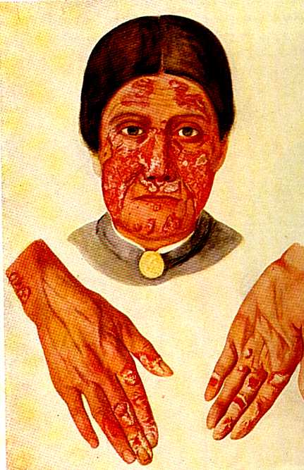
"In some circumstances (lupus) manifests itself at first as a violet rubefaction on this or that part of the face, and mainly on the nose, which at the same time is rather swollen: over many months the colour rises little by little; the surface becomes animated; a small ulcer forms and on top of it, a scab, which then thickens and covers the ulcer, which becomes progressively deeper. Lastly, the skin may get thinner in imperceptible stages and adopt the appearence of a scar, without there being tubercles or ulcers, and without displaying worse injuries than a livid colour and, from time to time, a light and barely perceptible peeling"10.
Cazenave in his book "Abreg� pratique des maladies de la peau" faithfully adhered to the classification that Biett had proposed11. However he introduced some new orders of diseases. Amongst them were lupus. After a few years, Ferdinand von Hebra (1816-1880) accepted the existence of the illness under the name lupus erithematosus 12,13.
The frequency of tuberculosis at that time was considerable. In the same five years when Kaposi diagnosed 22 cases of DLE, 279 cases of lupus vulgaris were seen in the same department16. One of his female patients with discoidal lupus erythematosus died displaying pulmonary tuberculosis. In spite of the extraordinary frequency of tuberculosis at that time, and that lupus and cutaneous tuberculosis were then considered related affections, Kaposi maintained that discoidal lupus erythematosus had no relation whatsoever to tuberculosis. Not everyone supported this opinion, and it met with special resistence amongst French dermatologists, and was disputed until beyond the first third of the 20th century. Kaposi used the term disseminated lupus erythematosus to refer to those cases in which there are dispersed cutaneous injuries, rather than those which exhibit visceral affection. However, he described some cases which involved fever, pleuritis, arthralgias and arthritis.
William Osler (1849-1919), from 1894 to 1903, observed 29 patients who presented "erythema with visceral injuries". It is generally accepted that many of these cases corresponded to descriptions of lupus erythematosus, although it is likely that revising them under present criteria, not all Osler's cases could be labelled in this way. At any rate, it was Osler who first indicated a renal affection or one affecting the central nervous system in cases of lupus erythematosus18,19.
A little later, in 1908, Alfred Kraus and Carl Bohac described the possible pulmonary affection of the syndrome: lupic pneumonia20. From then onwards other new visceral manifestations were described and this completed the clinical description of disseminated lupus erythematosus. In 1923, Emanuel Libman (1872-1946) and Benjamin Sacks contributed four cases of non-infectious endocarditis. Three of them displayed cutaneous injuries of LE21. The skin alterations were lacking in some cases, which caused the authors to hesitate in ascribing endocarditis to the LE table. In those times the greater part of studies of LE had been carried out by dermatologists, and perhaps for this reason the existence of cutaneous affection was considered essential for the diagnosis of lupus. Some years later, in 1936, George Belote and H.S. Ratner ratified that the endocarditis of Libman-Sacks was a manifestation of LE, even without the skin injuries22. A little later, in 1939, leukopenia and hypersensitivity to sunlight were described in cases of LE.
Discoidal lupus erythematosus and systemic lupus erythematosus for a long time were considered different diseases with nothing in common. The idea that they were forms of the same disease, and that they could even present transitional forms was introduced in 1937 by Harry Keil, although it must be said that quite a long time passed before this idea gained a general acceptance. It was also Keil who clearly outlined the differential diagnosis of cutaneous manifestations in order to distinguish between lupus erythematosus and dermatomyositis23.
For a long time lupus was considered a strange entity of very low incidence. Thus, only eight cases were diagnosed between 1897 and 1908 in the University-Hospital of Prague; three in the Johns Hopkins Hospital between 1919 and 1923; and five in the Mayo Clinic between 1918 and 192124. The causes of the low incidence of this disease were probably the ignorance of the entirety of its indications and the exclusively clinical diagnosis, based particularly on the cutaneous injuries. In effect, as the indications became better known an increased incidence is observed. In the Mayo Clinic, for example, the cases diagnosed between 1938 and 1947 rose to 132, a figure totally out of step with previous statistics25.
In 1954 it was discovered that LE cells formed as a consequence of the existence of a serous factor and the leucocytic nucleus. In 1958, Friou was able to develop a laboratory test which made it possible to quantify the antigen-antibody reaction by using fluorescent antihuman globulin. The determination of antinuclear antibodies (ANA) gradually established itself as the principal diagnostic test for SLE and also made possible the evaluation of the severity of the disease. However, this development was not entirely beneficial. It was soon discovered that there could be false positive indications, in other words, positive reactions in persons not having SLE. The false positive indications could be caused by various circumstances, such as other diseases of the connective tissue, or treatment with certain drugs. Also, in Africa, false positives were discovered in some cases of malaria. However, other tests for the detection of other antibodies continued to appear and this increased the reliability of laboratory diagnosis.
Some years before, in 1929, Philip S. Hench (1896-1965) had already observed that some arthritic patients could experience a temporary remission of their symptoms during pregnancy or coinciding with jaundice. Hench attributed this phenomenom to the existence of an adrenocortical hormone. Hench, like Kendall, worked in the Mayo Clinic and for this reason, in 1949, when cortisone became available to him in sufficient quantity he was the first who was able to prove its anti-inflammatory effects on rheumatoid arthritis. Fortunately, he used the correct dosage and the result was spectacular. A little later the effects of these substances and of corticotrophine on patients with LE were tested, with success. Soon the works of Hench30 aroused a lively interest in the whole medical establishment. Proof of the high expectations created is the fact that only a year later Reichtein, Kendall and Hench were awarded the Nobel Prize (1950).
Soon a derivative of cortisone, prednisone, synthesized in 1955, became the most common treatment for SLE31. Only some cases of renal and cutaneous affection resisted this treatment.
Discoidal LE also benefited from treatment with topical corticosteroids. In 1952 Sulzberger and Witten managed to produce a steroid, which they called compound F, later becoming known as hydrocortisone, which was active applied topically32. Subsequently, a great number of corticosteroid molecules of varying strength have been synthesized. Many of them have contributed to the treatment of LE.
Treatment with antimalarials began in 1951 with quinacrine, which was administered in cases of discoidal lupus erythematosus33. A little later, other antimalarials were used, such as chloroquine and hydroxichloroquine, which are not only useful on cutaneous forms but also have applications in certain cases of SLE.
In the treatment of lupus immunosupressive drugs have played an important part. The first of these was nitrogenous mustard, tried out for the first time in 1952. Subsequently azatioprine and ciclofosfamide were introduced34, whose main indication are cases of serious renal affection resistent to corticotherapy.
With this therapeutic potential (to which we should add the possibility of treating potentially fatal bacterial infections with increasingly effective antibiotics) the mortality rate from LE has substantially decreased. At present, the survival rate 15 years after diagnosis has reached an average of 75% of all cases, although there are differences to consider regarding sex, age and race35. It is to be hoped that current research improves still further the diagnostic and therapeutic prospects of this disease in the near future.